The simple answer to this question is 3 weeks and 1 day. The longer answer is a lot more interesting. Did you know, for example, that the heart beats about 54 million times between fertilization and the birth of the baby? Meanwhile, over your 80 odd years on this planet, your heart will beat approximately 3.2 billion times. But let’s learn a little more about the development of the heart on the fetus.
For many women, knowing they’re going to have a baby is an exciting experience. They typically first know this by seeing an ultrasound of the baby in the womb. The image usually includes a little heart. That heart and its rhythmic palpitations is very important to the wellbeing of your baby. Modern medical technology has made it possible for doctors to collect important information about the baby’s heartbeat and even detect any congenital heart conditions at a very early stage.
The Development of the Fetal Heartbeat
 Source: https://www.whattoexpect.com
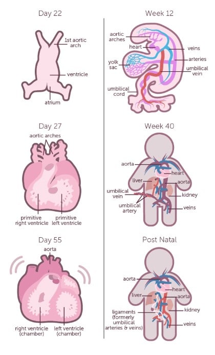
The heart typically starts out tube-shaped and gradually develops chambers and other features over time so it looks like a normal heart by the time the baby is born.
By the third week, or on the 22nd day, the fetal heart has begun to form. This is precisely when it begins to beat. However, the beat is too soft to be heard by heard through conventional stethoscopes. If the doctor really needs to hear the heartbeat, they can use the much more sensitive fetoscope. It can still be a challenge to hear the heartbeat though, with all the internal noises in the mother’s body, and also I the fetus is active.
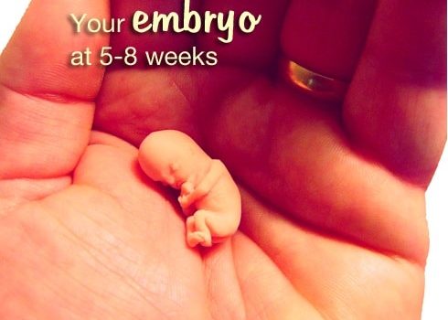
The heart chambers begin to develop by the 5th week. The heartbeat is still pretty soft at this point, but a lot easier to distinguish using specialized equipment.
In the 6th week the heart rate increases to the range between 100 beats per minute and 160 beats per minute. At this stage, the heart can be seen in ultrasound monitors beating.
By the 8th week, the heart has gained a steady rhythm, though it still beats much faster than the mother’s own heart.
The heart rate increases to a maximum of about 170 beats per minute in the 10th week, before slowing down and stabilizing around 130 beats per minute at the time of birth.
No one really knows how the heart begins beating, or what powers it, or even what controls the rhythm, but scientists agree that the answers to these questions could help us understand heart defects better and develop more effective treatments.
The Devices used to Measure the Fetal Heartbeat
The fetal heartbeat is measured using different equipment, each appropriate at a different developmental stage and designed to measure a different aspect of the heartbeat. This could either be the rhythm or the heart rate.
The fetal Doppler uses sound waves to map internal organs. It is moved on the surface of the mother’s womb and sends sound waves into the body. These sound waves bounce off the internal organs and form patterns on a screen. The fetal Doppler does not display any images, however.
After the 6th week of pregnancy, a transvaginal ultrasound can be used. This method involves inserting a probe into the mother’s vagina, which then sends sound waves bouncing off of the walls of the uterus. These rebounding waves are then converted into images of the different internal organs of the fetus, including the heart, from which the heartbeat can be monitored, and the progress of pregnancy can be determined.
The most accurate method to monitor the heartbeat is used just prior to birth. Just after the amniotic sac breaks and the amniotic fluid is released, an electrode is inserted into the mother’s cervix, attached to the baby’s head, and connected to a monitor. This monitor then allows the doctor to track both the baby’s heart rate and the uterine contractions of the mother.